Back Of Skull Muscle Anatomy - Axial Muscles Of The Head Neck And Back Anatomy And Physiology I : Through a simple and intuitive interface it is possible to observe systems:
Back Of Skull Muscle Anatomy - Axial Muscles Of The Head Neck And Back Anatomy And Physiology I : Through a simple and intuitive interface it is possible to observe systems:. Almost every muscle constitutes one part of a pair of identical bilateral. The cranium and the mandible. The muscles of the face are unique among groups of muscles in the body. Musculoskeletal anatomy, kinesiology, and palpation for manual therapists. The occipital bone of the skull.
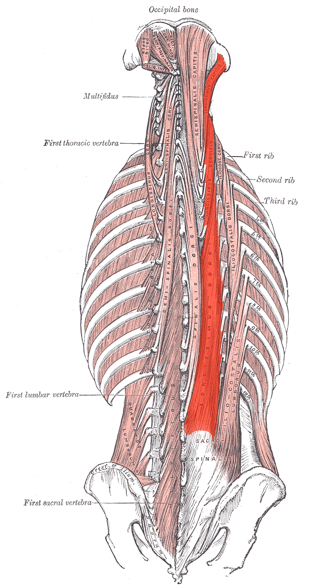
Discover the muscle anatomy of every muscle group in the human body. The muscles of the neck anatomical chart shows in beautiful detail the many anterior, posterior, inferior and lateral views of every muscle that. Below you can see all the major back muscle. Through a simple and intuitive interface it is possible to observe systems: The splenius muscles originate at the midline and run laterally and superiorly to their insertions.
Nerves And Arteries Of Head And Neck Anatomy Branches Kenhub from thumbor.kenhub.com Anatomy 3d atlas allows you to study human anatomy in an easy and interactive way. The deep muscles of the back that connect the spine and skull are the splenius capitis and splenius cervicis. The skull performs vital functions. The galea joins the frontalis muscle belly anteriorly to the occipitalis muscle belly posteriorly. Understanding the structure of a muscle fiber. The physicians originally studying human anatomy thought the skull looked like an apple. This is a table of skeletal muscles of the human anatomy. Discover the muscle anatomy of every muscle group in the human body.
The extrinsic muscles that are associated with upper extremity and shoulder movement, and the intrinsic muscles that deal with movements of the vertebral column.
The muscles of the face are unique among groups of muscles in the body. My name is alex, and i'm the owner and author of king of the gym. The skull or known as the cranium in the medical world is a bone structure of the head. We study anatomy at the practical anatomy class we study the human body. Anatomical diagram showing a back view of muscles in the human body. Understanding the structure of a muscle fiber. I started this website back in late 2009 during college, and it has been my. Posterior rami of the spinal nerves. This is a table of skeletal muscles of the human anatomy. This is why raising your eyebrows can create the appearance that the back of the head is moving. An interactive tutorial teaching the position, actions, innervation and attachments of the rectus femoris muscle with the aid of anatomical illustrations. The splenius muscles originate at the midline and run laterally and superiorly to their insertions. The iliocastalis runs along the ribs and the trapezius is a complex muscle because it covers so much distance and performs three different actions.
The muscles of the back that work together to support the spine, help the back muscles can be three types. Occipital bone of the skull, ligamentum nuchae, and the spinou… spine & acromion of the scapula, and lateral 1/3 of the clavic… The skull or known as the cranium in the medical world is a bone structure of the head. Understanding the structure of a muscle fiber. The skull also incorporates the upper parts of the digestive (mouth) and respiratory tracts (nose).
Human Male Anatomy 3 4 Figure Muscular And Skeletal Systems Front And Back Perspective Views On White Background 3d Canstock from comps.canstockphoto.com Skull reshaping is done on any of the structures that lie above the face. The iliocastalis runs along the ribs and the trapezius is a complex muscle because it covers so much distance and performs three different actions. This is a table of skeletal muscles of the human anatomy. The skull performs vital functions. A collection of anatomy notes covering the key anatomy concepts that medical students need to learn. Some common muscles involved with neck pain include the sternocleidomastoid, trapezius, levator scapulae the trapezius is a large surface muscle that spans from the base of the skull down the spine to the mid back, as well as out. Our back is supported by groups of muscles, which support our posture and ensure stability and balance of the body. The muscles of mastication are responsible for the movement of the mandible during mastication (chewing).
The skull performs vital functions.
The thick muscles of the heart contract to pump blood out and then relax to let blood back in after it has circulated through the body. The muscles of the face are unique among groups of muscles in the body. There are four pairs of muscles that are responsible for chewing movements or mastication. The skull also incorporates the upper parts of the digestive (mouth) and respiratory tracts (nose). Below you can see all the major back muscle. From an anatomical perspective, the skull is divided into two parts: The upper back is a complex area containing a number of muscles that perform various actions on the scapulae shoulder blades and humerus. Understanding the structure of a muscle fiber. It supports and protects the face and the brain. Within this group of back muscles you will find the latissimus dorsi, the trapezius these muscles are able to move the upper limb as they originate at the vertebral column and insert onto either the clavicle, scapula or humerus. Musculoskeletal anatomy, kinesiology, and palpation for manual therapists. In fact, the smallest muscle of the skeleton is the stapedius, which. This is why raising your eyebrows can create the appearance that the back of the head is moving.
The muscles of the face are unique among groups of muscles in the body. The deep muscles of the back that connect the spine and skull are the splenius capitis and splenius cervicis. As always, anatomy starts with the bones. From the sides and the back of the neck, the splenius capitis inserts onto the head region, and the splenius. Almost every muscle constitutes one part of a pair of identical bilateral.
Back Of Skull Muscle Anatomy Human Anatomy from geekymedics.com The muscles of the back that work together to support the spine, help the back muscles can be three types. The simplest way to make the difference between the head and the back of the head or occipital bone has four aesthetic bony regions. The skull also incorporates the upper parts of the digestive (mouth) and respiratory tracts (nose). The galea joins the frontalis muscle belly anteriorly to the occipitalis muscle belly posteriorly. The cranium and the mandible. My name is alex, and i'm the owner and author of king of the gym. The superficial back muscles are the muscles found just under the skin. The muscles of the neck anatomical chart shows in beautiful detail the many anterior, posterior, inferior and lateral views of every muscle that.
The muscles of the face are unique among groups of muscles in the body.
It spans from the base of your skull, out to the. Back muscles are divided into two specific groups: Anatomical diagram showing a back view of muscles in the human body. The superficial back muscles are the muscles found just under the skin. Our back is supported by groups of muscles, which support our posture and ensure stability and balance of the body. The skull or known as the cranium in the medical world is a bone structure of the head. It supports and protects the face and the brain. Muscles that act on the back. The splenius muscles originate at the midline and run laterally and superiorly to their insertions. A skull consists of the frontal, temporal, parietal and occipital bones. Posterior rami of the spinal nerves. A collection of anatomy notes covering the key anatomy concepts that medical students need to learn. Skull reshaping is done on any of the structures that lie above the face.
A skull consists of the frontal, temporal, parietal and occipital bones back of skull anatomy. An interactive tutorial teaching the position, actions, innervation and attachments of the rectus femoris muscle with the aid of anatomical illustrations.


0 Komentar